AG Wintergerst

Priv.-Doz. Dr. med. Maximilian W. M. Wintergerst, FEBO
Research areas
PD Dr. Wintergerst’s research group is dedicated to exploring innovative ophthalmic imaging approaches and their applications for international ophthalmology and uveitis. Dr. Wintergerst has published over 86 scientific papers (thereof 34 first- / last-authorships) and raised more than 2.000.000 € in funding. Among different international projects that he is heading, he is director of the Hospital Partnership Program of the University Hospital Bonn and the Sankara Eye Hospital Bangalore, India.
Dr. Wintergerst’s interest in imaging methods and image analysis began with his doctoral thesis in basic immunological science where he established a novel imaging and analysis method for cell culture at LMU Munich. Continuing this interest, he established his research focus in ophthalmic imaging and image analysis at University Hospital Bonn and completed his habilitation thesis on the evaluation of Smartphone-based retinal imaging for ophthalmologic examination at the University of Bonn.
Uveitis
Uveitis is a rare intraocular inflammation, especially affecting younger population, often resulting in significant visual impairment.
The absence of standardized nomenclature and assessment methods for intraocular inflammation contribute to a limited evidence base for therapeutic approaches. Consequently, defining clinical endpoints in research settings becomes notably challenging.
While the “Standardization of Uveitis Nomenclature” (SUN) working group has enhanced nomenclature, the evaluation of intraocular inflammation remains heterogenic and lacks reproducibility.
Focus on finding reliable endpoints to quantify intraocular inflammation using novel imaging techniques and automated image analysis
- Establishment of new qualitative and quantitative prognostic biomarkers for uveitis intermedia, posterior and panuveitis
- Validation of objectifiable readouts for intraocular inflammation suitable as a structural clinical endpoint for intervention studies
- Multimodal approach using non-invasive imaging methods
- OCT-Angiography
- enables high-resolution visualization of retinal and choriocapillary perfusion and analysis of the effects of inflammation on the microcirculation
- OCT-Angiography
- Anterion -OCT
- enables high-resolution visualization of the anterior segment - currently we’re probing the device for imaging of inflammatory cells in the anterior chamber and anterior vitreous
- Anterion -OCT
- Multimodal Fundusautofluorescence (FAF)
- promising approach in the diagnosis and monitoring of posterior uveitis and panuveitis, as changes in the FAF can be used to assess disease activity not visible by funduscopy or conventional color fundus photography
- Multimodal Fundusautofluorescence (FAF)
- Slit-Lamp Imaging
- color photography represents an optimal interface between conventional slit-lamp examination and novel imaging methods
- Slit-Lamp Imaging
International ophthalmology
Access to eye care professionals and, consequently, adequate eye care, remains a challenge in low- and middle-income countries (LMICs). In its Vision 2020 Action Plan, the World Health Organization (WHO) identified Diabetic Retinopathy (DR) as a priority eye disease, being the leading cause of visual impairment among working-age adults, affecting one-third of individuals with diabetes worldwide. Early detection of DR and other related conditions is imperative in reducing the prevalence of blindness. However, under resourced healthcare systems and notably deficient healthcare services in rural areas pose significant barriers. Therefore, the University Eye Hospital Bonn has initiated several collaborations with partners in LMICs, aiming to establish smartphone-based telemedicine screening and other projects to support and enhance healthcare services in these countries.
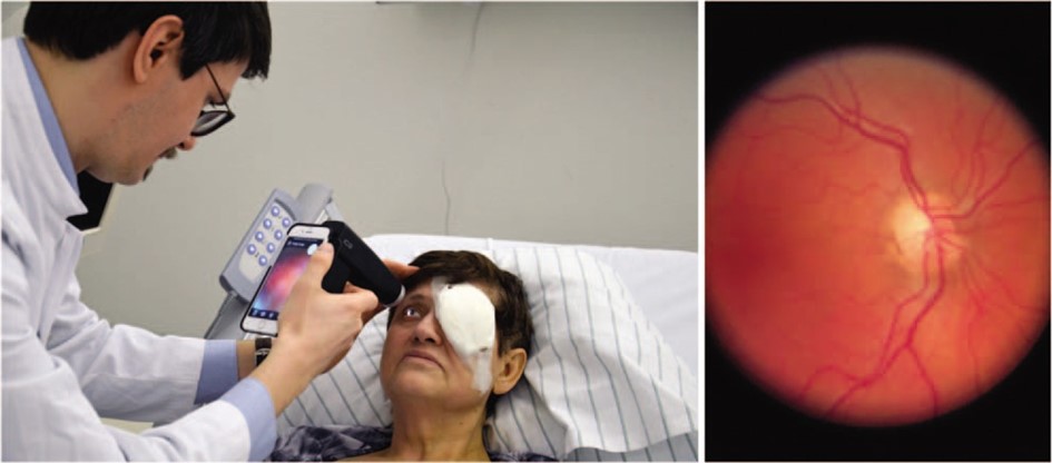
Smartphone-based fundus imaging (SBFI)
SBFI can be widely used for the detection of several eye diseases of the fundus with the help of medical assistants, who transfer captured images to telemedic grading centers. This method brings out its benefit in underserved rural or urban areas, where it improves the eye care access and lowers the barrier for less privileged populations to undergo a medical examination. The main focus of our SBFI approach as of right know is Diabetic retinopathy (DR). An extension of this screening towards glaucoma, retinopathy of the premature, and other retinal diseases is planned and its feasibility has been shown.
Artificial intelligence (AI)
In collaboration with Microsoft and the Sankara Eye Foundation in Bangalore, India, we are developing artificial intelligence algorithms with a low-cost approach in association with smartphone-based image capturing in order to improve cataract surgery outcomes in LMIC with the help of AI powered direct response teaching systems. Additionally, in cooperation with the University of Maastricht, the University of Bonn computing department, and the Sankara Eye Foundation, we are developing AI algorithm for automated retinal imaging analysis, which allow to support grading ophthalmologist to improve the efficacy, as well as It gives the opportunity of immediate orientating diagnostic response for patients in screening areas, where no doctor is present. The feasibility for Diabetic retinopathy as well as glaucoma screening has been demonstrated.
The simplicity of SBFI
The SBFI principle of scaling is to teach medical aid personnel in SBFI with the help either of an e-Learning class based on the University of Bonn e-learning platform, which can be hosted locally on each project partners server and has shown good results in medical aid personnel teaching. Alternatively, in-person teaching is employed, where local doctors are instructed in SBFI to disseminate knowledge locally to their medical aid personnel. These personnel then conduct screening camps in hard-to-reach rural or urban areas without proper medical healthcare service access.
Medical assistants then captures images in vision camps using SBFI, which are then uploaded into the digital patient health record app. Within this app, results can be retrieved and telemedic doctors can grade those captured images.
Patients finally get results in which time period they may need a checkup or whether they should travel to the closest hospital for treatment if necessary.
International Collaboration Partners
We are proud of our cooperation with several namely institutions worldwide mainly in Low - middle Income Countries (LMIC).
India
Since 2016, we have successfully partnered with the Sankara Eye Hospital in Bangalore and the Sankara Eye Foundation, led by Dr. Kaushik, in telemedical smartphone-based DR Screening. Together, we have established several vision camps, as well as our the Sudarshan Eye Sight app. Presently, we are continuing this work and have extended our collaboration to our new cataract surgery improvement project.
Bangladesh
For several years, the ORCD Bangladesh, an organization dedicated to rural development and the University of Bonn have collaborated on several projects to enhance eye health in Bangladesh. Starting in 2022, we initiated the implementation of our SBFI approach in Bangladesh to further improve access to eye healthcare in Bangladesh.
Nigeria
Since 2022, we have started our Collaboration with the University of Calabar Teaching Hospital in Nigeria, where we are currently jointly establishing a SBFI DR Screening approach.
Ghana
Additionally, since 2022, we have begun a partnership with the University of Cape Coast in Ghana, where we have so far dedicated our work towards research on the adaptability of our SBFI approach to different smartphones. We aim to broaden our approach here and implement regular DR screenings based on our collaboratively established SBFI approach.
since 07/2023 | Research group leader and Senior Researcher in Ophthalmology, University Hospital Bonn |
10/2022 | Fellow of the European Board of Ophthalmology (FEBO) |
since 06/2022 | University lecturer in Ophthalmology (Privatdozent) Bonn University |
06/2022 | Habilitation (highest German university degree) in Ophthalmology at the University of Bonn, Germany Habilitation thesis: „Evaluation of Smartphone-based fundus imaging for ophthalmologic examination” |
04/2021 - 06/2023 | Consultant Ophthalmologist University Hospital Bonn Clinical focus: retina and uveitis, cataract and vitreoretinal surgery |
Since 02/2019 | Director Hospital Partnership University Hospital Bonn - Sankara Eye Hospital Bangalore, India |
2017 | Dissertation (Dr. med.): summa cum laude (with highest honor) |
2015 - 2021 | Resident in Ophthalmology University Hospital Bonn (Prof. Dr. med. Frank G. Holz) |
2010 - 2015 | Medical Degree (M.D.), German state examination: 1.0 (top grade) Ludwig-Maximilians-Universität (LMU) Munich, Germany University of Cincinnati, USA Weill Cornell Medical College New York, USA |
2011 - 2013 | Doctoral studies within the academic course „Molecular medicine”, LMU Munich |
2008 - 2010 | Preclinical studies: German state examination: 1.0 (top grade) LMU Munich and Technical University of Munich, Germany |
Priv.-Doz. Dr. med. Maximilian W. M. Wintergerst, MD, FEBO (group leader)
Dr. med. Marie Just, MD
Raphael Lechtenböhmer, MD
Julie Jungblut, MD
Simon Müller, M.Sc. in Computer Science
Freya Behrens, B.Sc. (Master student, WHF)
Johanna Heidel (medical student, SHK)
Simon Jansen (medical PhD student)
Alexandra Weigandt (medical PhD student)
Selina Foti, B.Sc. (Master student, WHF)
Finn V. Hille (medical PhD student)
Lucas P. Reindl (medical PhD student)
Former members:
Gabriela Guzman (Master student, WHF)
Ilkin Yunuszada, MD
Markus Zeller
Lennart Overbeck, MD
Jana Katharina Koch, MD
Dr. med. Nicholas Merten, MD
Dr. med. Linus Jansen, MD
66 publications in peer-reviewed Journals (including Ophthalmology, American Journal of Ophthalmology, Nature Scientific Reports, IOVS, Journal of Experimental Medicine, Annals of the Rheumatic Diseases), thereof 28 first- / last-authorships. H-index: 19.
All Publications:
Selected Publications:
Wintergerst MWM et al. "Diabetic retinopathy screening using smartphone-based fundus imaging in India." Ophthalmology, 2020, 127(11):1529-1538
Wintergerst MWM et al. "Optical coherence tomography angiography in intermediate uveitis." American Journal of Ophthalmology, 2018, 194:35-45
Wintergerst MWM et al. "Non-contact smartphone-based fundus imaging compared to conventional fundus imaging: a low-cost alternative for retinopathy of prematurity screening and documentation." Scientific Reports, 2019, 9(1):19711
Wintergerst MWM et al. “Undilated versus dilated monoscopic smartphone-based fundus photography for optic nerve head evaluation” Scientific reports 8 (1), 10228
Wintergerst MWM et al. „A novel device for smartphone-based fundus imaging and documentation in clinical practice: comparative image analysis study” JMIR mHealth and uHealth, 2020, 8(7):e17480
Wintergerst, Maximilian WM, et al. "Smartphone-based fundus imaging–Where are we now?"
Asia-Pacific Journal of Ophthalmology, 2020, 9(4):308-314
Wintergerst MWM* and Merten NR* et al. „Spectrally Resolved Autofluorescence Imaging in Posterior Uveitis.” Scientific Reports. 2022 Aug 29;12(1):14337
Wintergerst MWM et al. “Structural endpoints and outcome measures in uveitis” Ophthalmologica, 2021; 244(5):465-479
Wintergerst MWM et al. „Vessel density on optical coherence tomography angiography is prognostic for future disease course in intermediate uveitis.” Scientific reports. 2024 Feb 5;14(1):2933
Mauschitz MM*, Zeller M*, … , Wintergerst MWMW „Fundus Autofluorescence in Posterior and Panuveitis - An Under-Estimated Imaging Technique: A Review and Case Series” Biomolecules 2024, 14(5), 515